Brighton Implant Clinic - An award winning dental health clinic


18
Share
Root Canal Treatment
Dentist X-rays & Root Canal Treatment (RCT): What to Expect & Why It Matters
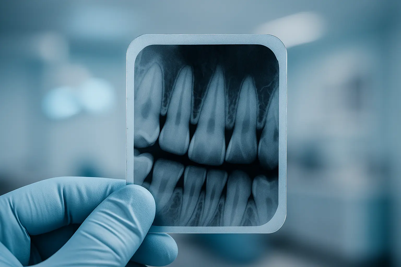
When it comes to root canal treatment (RCT), radiographs play an indispensable role in diagnosis, treatment planning, and monitoring. While commonly referred to as "X-rays," it's important to note that an X-ray is the technique used to produce a radiograph—the actual image of the tooth's internal structures.
Modern dentistry has largely transitioned from traditional film-based radiographs to digital formats, enhancing image clarity and reducing radiation exposure.
At Brighton Implant Clinic, we utilize advanced digital radiography to ensure precise diagnostics and effective treatment outcomes. Our commitment to cutting-edge technology allows us to provide patients with the highest standard of care in endodontic procedures.
The Science Behind Radiographs: How They Work

Radiographs are essential diagnostic tools that allow dentists to visualize the internal structures of teeth and surrounding bone. By directing controlled X-ray beams through the oral cavity, we capture detailed images that reveal issues not visible during a standard examination.
Digital radiography offers several advantages over traditional methods, including immediate image processing, enhanced image quality, and significantly reduced radiation exposure. These benefits contribute to more accurate diagnoses and streamlined treatment planning.
Safety is paramount in dental imaging. The radiation dose from dental radiographs is minimal, and protective measures such as lead aprons and thyroid collars are routinely used to safeguard patients. According to the Cleveland Clinic, dental X-rays are considered safe and are an integral part of comprehensive dental care.
Pre-operative Radiographs: Setting the Stage for Treatment
Before initiating root canal therapy, pre-operative radiographs are taken to assess the condition of the affected tooth. These images help identify the extent of decay, detect any abscesses or infections, and evaluate the tooth's anatomy.
The procedure involves positioning a digital sensor inside the patient's mouth, aligned with the tooth in question. The patient is asked to remain still while the X-ray is taken, ensuring a clear and accurate image.
Pre-operative radiographs are crucial for developing an effective treatment plan. They provide a baseline for comparison with subsequent images taken during and after the procedure, allowing for precise monitoring of treatment progress.
Intra-operative Radiographs: Guiding the Root Canal Procedure
During root canal treatment, intra-operative radiographs are utilized to guide the procedure and ensure its success. These images assist in determining the working length of the root canals, ensuring that the dentist cleans and shapes the canals to the appropriate depth.
Additionally, intra-operative radiographs help verify the removal of infected pulp tissue and confirm the proper placement of instruments within the canals. This step is vital to prevent complications and ensure the thorough cleaning of the root canal system.
At Brighton Implant Clinic, we employ advanced endodontic equipment, including nickel-titanium rotary files and digital radiography, to enhance the precision and efficiency of root canal procedures.
Post-operative Radiographs: Confirming Treatment Success
After completing the root canal procedure, post-operative radiographs are taken to evaluate the quality of the treatment. These images confirm that the canals have been adequately filled and sealed with gutta-percha, a biocompatible material used to prevent reinfection.
Post-operative radiographs also serve as a reference for future evaluations, allowing dentists to monitor the tooth's healing process and detect any potential issues early on.
Ensuring the success of root canal therapy relies heavily on the accuracy of these post-treatment images. They provide both the dentist and patient with confidence in the long-term prognosis of the treated tooth.
Recall Radiographs: Long-term Monitoring and Maintenance

Recall radiographs are periodic images taken during follow-up appointments to monitor the treated tooth's condition over time. These radiographs help assess the healing of periapical tissues and ensure that the tooth remains free from infection or other complications.
The frequency of recall radiographs depends on the individual case and the dentist's assessment. Regular monitoring is essential to maintain oral health and address any issues promptly.
At Brighton Implant Clinic, we emphasize the importance of ongoing dental care and provide personalized recall schedules to support our patients' long-term oral health.
Types of Radiographs Used in Root Canal Treatment
Various types of radiographs are employed during root canal therapy, each serving a specific purpose:
- Periapical Radiographs: These images capture the entire tooth, from crown to root tip, and are essential for diagnosing root and surrounding bone conditions.
- Bitewing Radiographs: Primarily used to detect decay between teeth and assess bone levels, these are less common in root canal procedures but may be used for comprehensive evaluations.
- Cone Beam Computed Tomography (CBCT): This advanced imaging technique provides three-dimensional views of the tooth and surrounding structures, offering detailed information for complex cases.
The selection of radiograph type depends on the clinical situation and the level of detail required for accurate diagnosis and treatment planning.
Interpreting Radiographs: What Dentists Look For

Dentists analyze radiographs to identify various indicators of dental health or pathology:
- Dark Areas: These may signify decay, infection, or bone loss.
- Root Canal Anatomy: Understanding the number and curvature of canals is vital for effective cleaning and filling.
- Periapical Radiolucency: This indicates infection at the root tip, often necessitating root canal treatment.
Accurate interpretation of these images is crucial for developing an effective treatment plan and ensuring the long-term success of the procedure.
Patient Experience: What to Expect During Radiographic Procedures
Understanding what to expect during the radiographic process can significantly reduce patient anxiety. Initially, you'll be asked to remove any metallic objects, such as jewelry or glasses, which might interfere with the radiograph clarity. You'll then sit upright as the dentist gently places a digital sensor inside your mouth. This small device captures clear, detailed images with minimal discomfort.
Throughout this process, it's crucial to remain still for a few seconds as the radiograph is taken. Any movement could blur the image, potentially requiring the procedure to be repeated. Our experienced team at Brighton Implant Clinic ensures your comfort by clearly explaining each step and addressing any concerns you may have.
Digital radiography is swift, painless, and safe, minimizing exposure time and providing immediate results. Patients often express relief and confidence upon seeing real-time images, as it helps them better understand their dental conditions and the importance of the recommended treatment.
The Importance of Radiographs in Ensuring Treatment Efficacy
Radiographs are fundamental for achieving optimal outcomes in root canal treatment. They enable dentists to make informed decisions by precisely diagnosing dental conditions, assessing treatment progression, and ensuring comprehensive infection removal.
Moreover, accurate radiographic imaging reduces the risk of future complications, such as recurrent infections or incomplete healing, by ensuring thorough canal cleaning and filling. At Brighton Implant Clinic, we utilize radiographs strategically at various stages—before, during, and after treatment—to maximize success and patient satisfaction.
Ultimately, embracing radiographic technology supports long-term oral health, preserves natural teeth, and contributes significantly to maintaining an attractive, healthy smile for years to come.
FAQs about Dentist X-rays & Root Canal Treatment
Why are multiple radiographs necessary during root canal treatment?
Multiple radiographs ensure accurate diagnosis, precise monitoring during treatment, and verification of successful sealing and filling of the canals. Each radiograph helps confirm the efficacy of every procedural step.
Are dental radiographs safe?
Yes, modern digital radiographs emit significantly lower radiation compared to traditional methods, making them exceptionally safe. Additionally, dental professionals use protective measures, such as lead aprons and thyroid collars, further reducing any minimal risks.
Can I refuse radiographs during my dental treatment?
Patients have the right to refuse radiographs; however, doing so could hinder accurate diagnosis and effective treatment planning. It's advisable to discuss any concerns with your dentist to make an informed decision.
How do radiographs help in detecting root canal issues?
Radiographs provide detailed images of internal tooth structures, clearly revealing infections, decay, and anatomical complexities. They are essential in diagnosing problems that aren't visible during regular oral examinations.
What is the difference between traditional X-rays and digital radiographs?
Digital radiographs offer numerous advantages over traditional film X-rays, including reduced radiation exposure, instant image availability, improved clarity, and easier image storage and retrieval.
Conclusion: Why Radiographs are Crucial to Your Dental Health
Radiographs are not merely supplemental—they are essential components of successful root canal therapy. By providing clear and detailed insights into tooth structure and pathology, radiographs help ensure treatments are accurate, thorough, and effective. At Brighton Implant Clinic, our dedicated dental professionals leverage advanced digital radiography to enhance treatment precision, ensuring your dental health and comfort every step of the way.
For more information about radiographic imaging, root canal treatment, or other dental procedures, please contact Brighton Implant Clinic or call us directly at 0800 111 6623. Your journey toward a healthier smile begins with informed, personalized care.