Brighton Implant Clinic - An award winning dental health clinic


18
Share
Root Canal Treatments
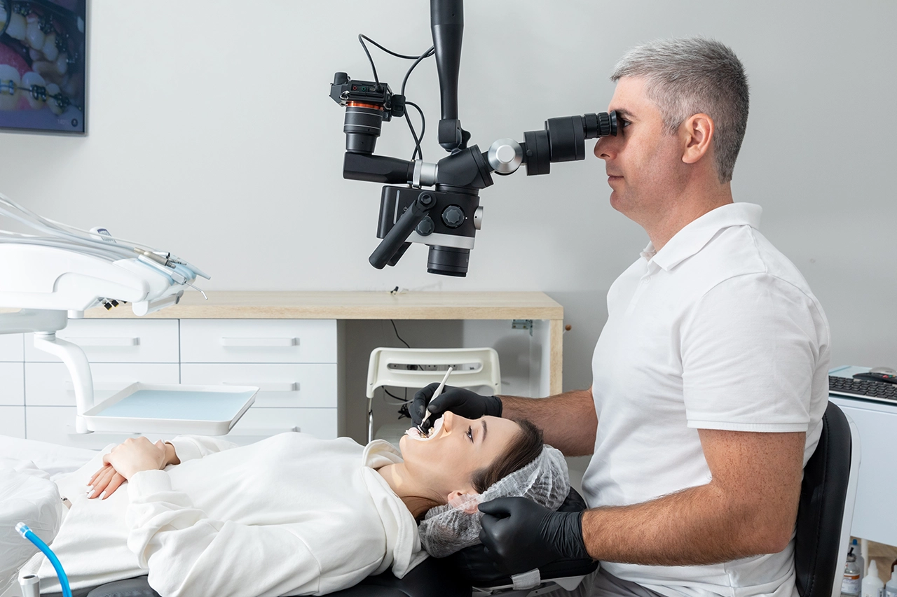
New Dentist & High-Powered Microscope For Root Canal Precision in Worthing
Root canal treatments have long been a cornerstone of restorative dentistry, essential for preserving natural teeth compromised by infection or trauma. However, the complexity of root canal systems often poses challenges, even for experienced practitioners. Advancements in dental technology, particularly the introduction of high-powered microscopes, have revolutionized the field, enabling unprecedented precision in endodontic procedures.
At Brighton Implant Clinic, we are committed to integrating cutting-edge technology to enhance patient outcomes. Our Worthing branch has recently welcomed Dr. Vanderley Da Silva, a seasoned endodontist with over a decade of experience. Complementing his expertise, we've invested in state-of-the-art dental microscopes, ensuring our patients receive the highest standard of care in root canal treatments.
Understanding Root Canal Treatment
To appreciate the advancements brought by high-powered microscopes in endodontic procedures, it's essential first to understand the fundamentals of root canal treatment.
What is a Root Canal?
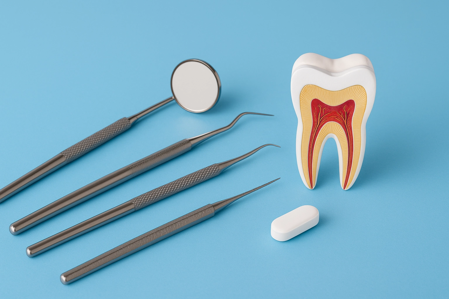
A root canal is a dental procedure aimed at treating infection at the center of a tooth (the root canal system). It involves removing the infected or damaged pulp, cleaning and disinfecting the canal, and then filling and sealing it to prevent further infection. This treatment allows patients to preserve their natural teeth, avoiding the need for extraction.
Common Causes for Treatment
Root canal treatments become necessary when:
- Tooth decay penetrates deep into the tooth, reaching the pulp.
- Repeated dental procedures on a tooth cause pulp inflammation.
- Cracks or chips in the tooth expose the pulp to bacteria.
- Trauma to the tooth leads to pulp damage, even without visible cracks.
These conditions can lead to severe pain and, if left untreated, may result in abscess formation or tooth loss.
Traditional Challenges
Historically, root canal treatments relied heavily on tactile sensation and two-dimensional radiographs. This approach often made it difficult to detect all canals, especially in teeth with complex anatomies. Missed canals or incomplete cleaning could lead to persistent infections or the need for retreatment.
The Role of High-Powered Microscopes in Dentistry
With a foundational understanding of root canal treatments, we can now explore how high-powered microscopes have transformed the field of endodontics.
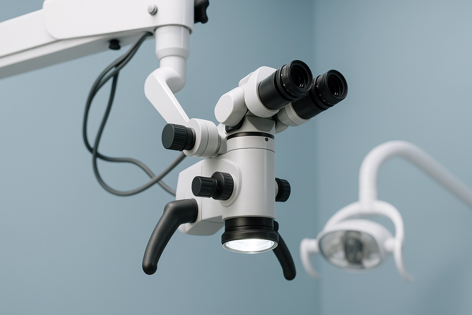
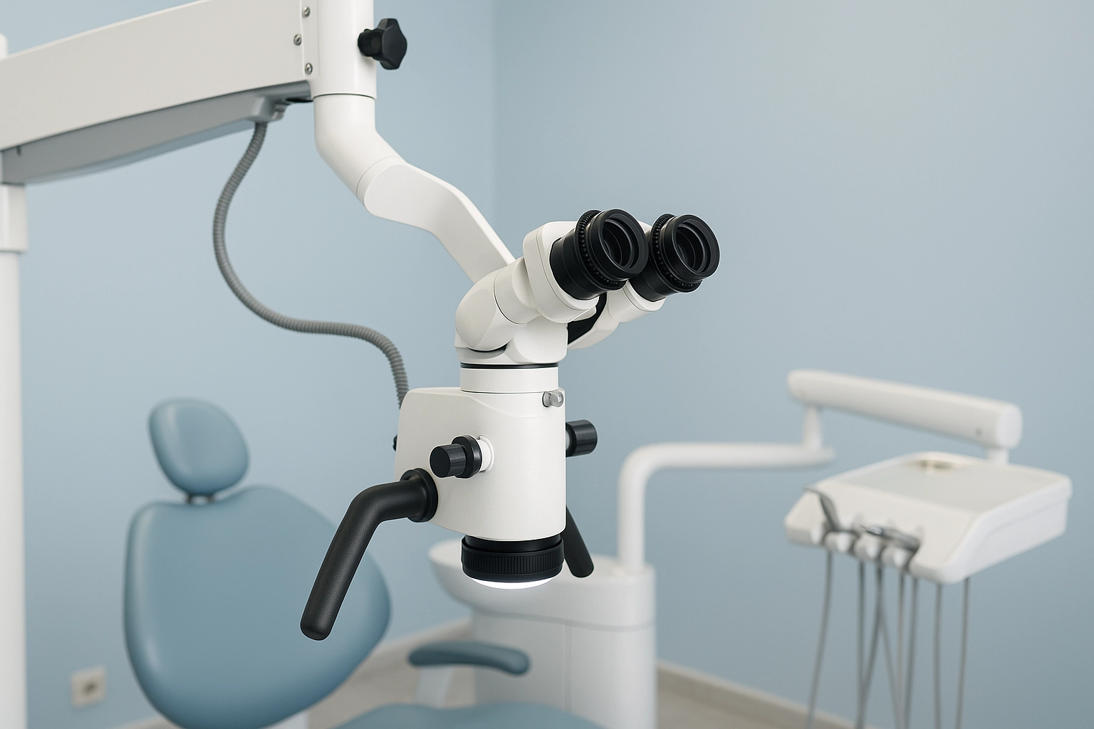
The advent of dental operating microscopes has transformed endodontic practices. These microscopes provide magnification levels ranging from 4x to 25x, allowing dentists to visualize the intricate details of the tooth's internal structure.
Enhanced illumination further aids in identifying and treating complex canal systems.
Magnification Benefits
High magnification enables dentists to:
- Identify additional canals that might be missed with the naked eye.
- Detect microfractures or calcifications within the tooth.
- Ensure complete removal of pulp tissue and bacteria.
- Perform procedures with greater accuracy, preserving more of the natural tooth structure.
Illumination Advantages
Modern dental microscopes are equipped with coaxial illumination, providing shadow-free lighting directly into the canal system. This feature is crucial for:
- Visualizing the full depth of the canal.
- Ensuring thorough cleaning and shaping.
- Reducing the risk of leaving behind infected tissue.
Advantages of Microscope-Assisted Root Canals
Building upon the technological capabilities of dental microscopes, let's delve into the specific benefits they offer in root canal procedures.
Enhanced Precision
Microscope-assisted root canals allow for meticulous cleaning and shaping of the canal system. The increased visibility ensures that even the most intricate canal anatomies are thoroughly treated, reducing the risk of reinfection.
Detection of Microfractures
Microfractures, often invisible to the naked eye, can compromise the success of a root canal treatment. With high-powered microscopes, dentists can identify these fractures early, allowing for appropriate interventions and increasing the longevity of the tooth.
Minimally Invasive Approach
The precision offered by microscopes means that dentists can remove only the necessary amount of tooth structure, preserving as much of the natural tooth as possible. This conservative approach promotes better structural integrity and function post-treatment.
Improved Success Rates
Studies have shown that the use of dental operating microscopes in endodontic procedures leads to higher success rates and reduced need for retreatments. The enhanced visualization ensures comprehensive treatment, minimizing the chances of missed canals or residual infection.
Dr. Vanderley Da Silva: Expertise in Endodontics
Delivering advanced dental care requires not only technology but also exceptional clinical skill. At Brighton Implant Clinic, Dr. Vanderley Da Silva leads our endodontic treatments with a unique blend of expertise, precision, and patient-focused care.
Professional Background
Dr. Da Silva earned his dental degree from the Federal University of Minas Gerais (UFMG) and specialized in endodontics at the University of São Paulo (USP) in Bauru, Brazil. He continues to expand his expertise through ongoing postgraduate training under internationally recognized experts like Professor Gilberto Debelian.
Experience and Skills
With over 20 years of experience, Dr. Da Silva has managed a wide range of complex root canal cases. His proficiency with high-powered dental microscopes allows for enhanced precision, accurate detection of hidden canals and fractures, and minimally invasive treatment that preserves tooth structure.
Patient-Centered Approach
Known for his calm and compassionate manner, Dr. Da Silva ensures patients feel informed and comfortable at every stage of their treatment. Fluent in English, Spanish, and Portuguese, he communicates clearly and effectively with patients from diverse backgrounds.
His presence strengthens Brighton Implant Clinic’s commitment to advanced, patient-first dental care.
Brighton Implant Clinic's Investment in Advanced Technology
To deliver exceptional dental care, it's essential to combine skilled professionals with state-of-the-art technology. At Brighton Implant Clinic, our commitment to excellence is reflected in our continuous investment in advanced equipment and training.
State-of-the-Art Equipment
Our clinics are equipped with cutting-edge dental operating microscopes, providing magnification up to 25x. This technology allows our dentists to visualize the minutiae of the oral cavity, ensuring precise diagnosis and treatment. The enhanced visualization capabilities of these microscopes facilitate the identification of complex canal anatomies and microfractures that might be missed with traditional methods.
Training and Expertise
We believe that technology is only as effective as the professionals who use it. Our dental team undergoes rigorous training to master the use of high-powered microscopes, ensuring that each procedure is performed with the utmost precision. This dedication to continuous learning ensures that our patients receive care that aligns with the latest advancements in dental science.
Clinic Locations
Accessibility to top-tier dental care is a priority for us. With multiple branches across East and West Sussex—including Brighton, Hove, Hailsham, and Worthing—patients can conveniently access our specialized services. Each location upholds our commitment to excellence, providing consistent, high-quality care across all clinics.
Step-by-Step: Microscope-Assisted Root Canal Procedure
Understanding the process of a microscope-assisted root canal can alleviate patient anxiety and highlight the benefits of this advanced approach. Here's a detailed walkthrough of what patients can expect during the procedure.
Initial Assessment
The journey begins with a comprehensive evaluation, including digital radiographs and, if necessary, cone-beam computed tomography (CBCT) scans. These diagnostic tools provide detailed images of the tooth's internal structures, allowing for accurate assessment of the infection's extent and the tooth's anatomy. This information is crucial for planning a precise and effective treatment strategy.
Access and Cleaning
Once the treatment plan is established, the dentist administers local anesthesia to ensure patient comfort. Using the dental operating microscope, a small access opening is created in the tooth to reach the infected pulp. The enhanced magnification and illumination allow for meticulous cleaning and shaping of the root canals, ensuring the removal of all infected tissue and debris.
Filling and Sealing
After thorough cleaning, the canals are disinfected and dried. They are then filled with a biocompatible material, such as gutta-percha, to seal the canals and prevent reinfection. The access opening is sealed with a temporary or permanent filling, depending on the treatment plan. The precision afforded by the microscope ensures that the filling material is placed accurately, promoting optimal healing.
Post-Treatment Care
Following the procedure, patients receive detailed aftercare instructions to support healing. A follow-up appointment may be scheduled to monitor progress and, if necessary, place a permanent restoration like a crown. The use of a microscope throughout the procedure enhances the accuracy of the treatment, leading to improved success rates and patient satisfaction.
Comparing Traditional and Microscope-Assisted Techniques
To fully appreciate the advancements in endodontic care, it's helpful to compare traditional root canal methods with microscope-assisted techniques. This comparison highlights the tangible benefits of incorporating high-powered microscopes into dental procedures.
The enhanced visualization and precision provided by the microscope lead to more thorough cleaning and sealing of the canals, reducing the risk of reinfection and the need for retreatment.
Frequently Asked Questions (FAQs)
What makes microscope-assisted root canals more effective than traditional methods?
Microscope-assisted root canal treatments offer enhanced magnification and illumination, allowing dentists to detect intricate canal structures and microfractures that might be missed with traditional methods. This precision ensures thorough cleaning and sealing of the canals, reducing the risk of reinfection and the need for retreatment.
Is the procedure more time-consuming with the use of a microscope?
While the initial setup may take slightly longer, the overall procedure can be more efficient due to the improved visualization and precision. This efficiency can potentially reduce the number of appointments required and lead to quicker recovery times.
Will I experience less pain with microscope-assisted treatment?
Yes, the precision of the procedure typically results in less tissue trauma, leading to reduced postoperative discomfort and faster recovery times. Patients often report a more comfortable experience compared to traditional root canal treatments.
Are all dentists trained to use high-powered microscopes?
Not all dentists have specialized training in using dental microscopes. At Brighton Implant Clinic, our team, including Dr. Vanderley Da Silva, is proficient in utilizing this technology to enhance patient outcomes.
How do I know if I need a microscope-assisted root canal?
A thorough examination and consultation at our clinic will determine the most appropriate treatment approach for your specific dental needs. If your tooth has complex anatomy or previous treatments have failed, microscope-assisted therapy may be recommended.
Conclusion
Microscope-assisted root canal therapy represents a significant advancement in endodontic treatment, offering enhanced precision, improved outcomes, and a more comfortable patient experience.
At Brighton Implant Clinic, our commitment to integrating cutting-edge technology, such as high-powered dental microscopes, ensures that our patients receive the highest standard of care. With the expertise of specialists like Dr. Vanderley Da Silva, we are equipped to handle even the most complex root canal cases with confidence and precision.
If you're experiencing dental discomfort or have been advised to undergo root canal therapy, consider the benefits of microscope-assisted treatment. Our experienced team is here to provide you with exceptional care tailored to your needs.